Dr. Phyto
bacterial blight / canker (multi-host)
Pseudomonas syringae pv. syringae
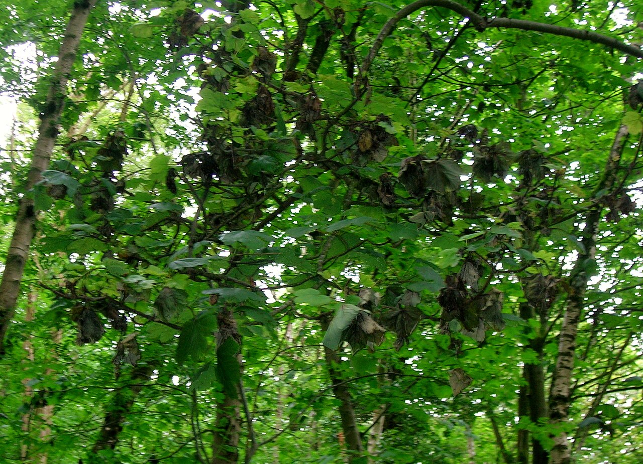
Symptoms
Brown-black angular leaf spots with yellow halos, blackened buds + young shoots that fail to open, sunken brown cankers on stems with bacterial ooze in spring, blossom blight in cool wet bloom periods, branch dieback in heavy infections.
Plants affected
Easily confused with
- sclerotinia stem rot
How to tell them apart: Pseudomonas syringae pv. syringae (bacterial blight) on Delphinium shows greasy, water-soaked dark-brown to black streaks and angular lesions on stems, leaves and flower stalks; lesions stay slimy/wet, often have a thin yellow halo on leaves, and may exude a milky bacterial ooze when cut and squeezed in a glass of water (a streaming 'bacterial thread'). There is NO cottony growth and NO hard black bodies. Symptoms spread fastest in cool, wet, splashing-rain weather. Sclerotinia sclerotiorum (white mould / crown rot) starts as a soft, watery brown rot at the stem base or crown, but quickly produces a conspicuous fluffy white cotton-wool mycelium over the rotting tissue, and inside the hollowed stem and on its surface forms hard, irregular black resting bodies (sclerotia) the size of rat droppings (2-10 mm). Stems bleach tan and become dry and shredded above the rot. The white fluff plus black sclerotia is diagnostic and never occurs with Pseudomonas; cut a suspect stem lengthwise and look for the black sclerotia inside.
- Phytophthora root + crown rot
How to tell them apart: On Photinia × fraseri, Phytophthora cinnamomi is a soilborne root and crown rot: the trouble starts below ground, so look for whole-branch or whole-plant wilting and dull-brown foliar collapse, sparse roots, and dark reddish-brown cambial staining when you cut the bark at the trunk base near the soil line; decline is worst in waterlogged, poorly drained soil. Pseudomonas syringae pv. syringae is an aerial bacterial disease: look up in the canopy for discrete angular reddish-purple to brown leaf spots, blackened sunken twig and shoot lesions, and cankers that girdle individual shoots from the tip downward, typically after cool wet spring weather or frost. Rule of thumb: Phytophthora kills from the roots up with collar staining, while Pseudomonas kills from the shoot tips down with distinct spots and twig cankers.
- entomosporium leaf spot
How to tell them apart: On Photinia × fraseri, Diplocarpon mespili (Entomosporium leaf spot) starts as tiny round bright-red to maroon spots scattered on both old and new leaves; as they age the centres turn grey-brown and develop a minute glossy black pinhead-sized fruiting body (acervulus) in the middle — use a hand lens to find this raised black dot, which is diagnostic and absent in bacterial disease. Pseudomonas syringae pv. syringae instead makes darker brown-to-black angular spots that are limited by the leaf veins, often water-soaked and translucent at the margin when held to the light, sometimes with a thin yellow halo, and appears mainly in cool wet spring weather on young shoots and leaf edges, frequently with twig dieback and no black fruiting dot. In short: look for the central glossy black speck and round red spots for Entomosporium, versus angular vein-limited water-soaked spots with shoot dieback for Pseudomonas.
- horse chestnut leaf blotch
How to tell them apart: On horse chestnut (Aesculus hippocastanum), bacterial blight (Pseudomonas syringae pv. syringae) shows small, water-soaked, angular dark-brown spots that are sharply confined by the leaf veins, often with a thin yellow halo, and the tissue may ooze or feel slightly slimy in wet weather; lesions appear scattered, including in the leaf interior, and can be linked to twig/bud cankers. Leaf blotch (Guignardia aesculi) instead begins as reddish-brown spots that enlarge into large, irregular, dry papery blotches typically starting at the leaf margin or tip, surrounded by a bright yellow border, with curled crispy edges and tiny black pinhead fruiting bodies (pycnidia) speckling the dead areas in late summer. If you see vein-limited angular wet spots, suspect the bacterium; if you see spreading brown papery blotches from the margins with black dots, suspect the fungus.
- Cucumber mosaic virus (CMV)
How to tell them apart: Cucumber mosaic virus produces a systemic, diffuse light-and-dark green mosaic, mottle and vein-clearing spread evenly across the whole wisteria leaf blade, with NO necrosis, water-soaking or sharp margins, and is often accompanied by puckering or narrowing of new leaflets. Pseudomonas syringae pv. syringae instead makes discrete spots that start water-soaked (greasy, translucent), then collapse into brown-to-black dead flecks surrounded by a distinct bright-yellow halo, are often angular because they are limited by the small veins, cluster toward leaf margins and tips, and appear after cool wet spring weather alongside sunken cankers and shoot dieback. The simplest field test: hold the leaf to the light — a soft chlorotic pattern with no dead tissue is the virus, whereas dead spots with greasy water-soaked edges and yellow rings are the bacterium.
- brown soft scale
How to tell them apart: Coccus hesperidum (brown soft scale) shows discrete, flattened oval, yellow-brown to amber bodies 3-4 mm long, firmly stuck and lined up along stems, petioles and the underside midribs of wisteria leaves; they are sticky to the touch and sit under a sooty, wipe-off black film of honeydew mould with ants often present, and the bark beneath stays sound. Pseudomonas syringae pv. syringae (bacterial blight/canker) instead produces fixed dark-brown to black sunken cankers and lesions in the bark and angular water-soaked brown leaf spots, often with a yellow halo; the damage is in the living tissue and cannot be scraped off, frequently oozes in spring and appears after cool wet weather rather than as raised insects. If you can lift a body off with a fingernail and the tissue beneath is clean, it is scale, not bacterial canker.
Treatment
Dr. Phyto builds a dated, step-by-step treatment plan with country-approved products — start a free diagnosis to get yours.
Common questions
- How do I recognise bacterial blight / canker (multi-host) on Japanese maple?
- Brown-black angular leaf spots with yellow halos, blackened buds + young shoots that fail to open, sunken brown cankers on stems with bacterial ooze in spring, blossom blight in cool wet bloom periods, branch dieback in heavy infections.
- Do I have to report bacterial blight / canker (multi-host)?
- No. bacterial blight / canker (multi-host) is not a notifiable organism, so there is nothing to report to an authority. Treat it as a normal garden problem.
- How is bacterial blight / canker (multi-host) treated?
- Mainly with biological and cultural measures — removing affected material, changing the conditions that favour it, and treating with agents approved in your country. The exact steps and their timing depend on the plant, the season and where you live: Dr. Phyto works them out in a free diagnosis.
Free first diagnosis · no sign-up to start